Ghada A Fawzy1,2 ![]() ,
Shagufta Perveen1,
Areej M Al-Taweel1,
Raha S Orfali1,
Lubna Iqbal3,
Mehreen Lateef4,
Rasool B Tareen5,
Shabana I Khan6
,
Shagufta Perveen1,
Areej M Al-Taweel1,
Raha S Orfali1,
Lubna Iqbal3,
Mehreen Lateef4,
Rasool B Tareen5,
Shabana I Khan6
For correspondence:- Ghada Fawzy Email: gzeineldin@outlook.com Tel:+966118050496
Received: 31 October 2016 Accepted: 20 January 2017 Published: 26 February 2017
Citation: Fawzy GA, Perveen S, Al-Taweel AM, Orfali RS, Iqbal L, Lateef M, et al. Evaluation of some biological activities of Abelia triflora R Br (Caprifoliaceae) constituents. Trop J Pharm Res 2017; 16(2):319-325 doi: 10.4314/tjpr.v16i2.9
© 2017 The authors.
This is an Open Access article that uses a funding model which does not charge readers or their institutions for access and distributed under the terms of the Creative Commons Attribution License (http://creativecommons.org/licenses/by/4.0) and the Budapest Open Access Initiative (http://www.budapestopenaccessinitiative.org/read), which permit unrestricted use, distribution, and reproduction in any medium, provided the original work is properly credited..
Purpose: To investigate the antioxidant, anti-inflammatory, antidiabetic, cardiovascular and cytotoxic activities of the leaf extract and major compounds isolated from Abelia triflora R. Br. (Caprifoliaceae)
Methods: The chloroform soluble fraction of A. triflora leaves was subjected to several column chromatographic separations to isolate its constituents. Anti-inflammatory and antioxidant activities were determined in terms of the ability to inhibit NF-kB, iNOS activity and lipoxygenase enzyme, and to decrease oxidative stress in HepG2 cells. Antidiabetic and cardiovascular activities were determined by screening for peroxisome proliferator-activated receptor alpha (PPARα) and PPAR=1; agonistic activities. In vitro cytotoxic activity was determined against a set of four human cancer cell lines (SK-MEL, KB, BT-549, SK-OV-3) and two non-cancerous kidney cell lines (LLC-PK1 and VERO). Cell viability was measured by neutral red assay.
Results: Three triterpene acids were isolated from the chloroform fraction namely; ursolic acid (4), 2, 3-dihydroxy ursolic acid (5) and 2, 3, 21-trihydroxy ursolic acid (6). The results showed that ursolic acid exhibited potent inhibition of lipoxygenase enzyme and iNOS (inducible nitric oxide synthase) activity with IC50 (half-maximal inhibitory concentration) value of 13.0 µg/mL, compared to parthenolide positive standard (IC50, 0.3µg/mL); furthermore, it inhibited NF-kB (nuclear factor-kappa B) with IC50 of 25.0 µg/mL, compared to parthenolide (positive standard, (IC50, 0.5 µg/mL). Also, ursolic acid possessed the highest cytotoxic effect against the three cell lines, SK-MEL (IC50, 14.5 µg/mL), BT-549 (IC50, 16.0 µg/mL) and SK-OV-3 (IC50, 12.5 µg/mL). Only 2,3-dihydroxy ursolic acid activated PPAR=1; (1.5-fold at 25 µM), compared to rosiglitazone (positive standard, 3.7 fold at 10 µM)
Conclusion: Among the investigated compounds, ursolic acid exhibited the highest anti-inflammatory and cytotoxic activities, while 2,3-dihydroxy ursolic acid demonstrated antidiabetic activity via activation of PPAR=1;.
Introduction
Genus Abelia (Caprifoliaceae) is composed of around eighty species found mostly in the Himalayas and East Asia. Abelia is represented by Abelia triflora in Pakistan and is available in Kaghan Valley (Hazara District) [1]. Little phytochemical work has been done on genus Abelia. Iridoid and bisiridoid glycosides have been isolated from Abelia grandiflora and Abelia chinensis [2-3]. Our previous investigation on A. triflora led to the isolation of five compounds from ethyl acetate and n-butanol fractions and their anticancer activities were explored [4].
The objective of the current study was to undertake a phytochemical investigation of the chloroform soluble fraction of the plant as well as the evaluation of the anti-inflammatory, antioxidant, cytotoxic, antidiabetic and cardiovascular activities of fractions and major isolated compounds
Methods
Plant material
Leaves of A. triflora (8.0 kg) were collected in March 2012, from Ziarat Valley, near Quetta, Baluchistan province, Pakistan and was identified by a plant taxonomist (Rasool Bakhsh Tareen), Department of Botany, Baluchistan University, Quetta; a voucher specimen (no. 300) was deposited in the herbarium of the department [4].
Extraction and isolation
The leaves were shade-dried, ground (8.0 kg) and extracted with methanol (3 × 10 L) at room temperature. The combined methanol extract was evaporated under reduced pressure to obtain a thick gummy residue (500 g). It was suspended in water and sequentially extracted with the following solvents to give fractions that weighed; n-hexane (120 g), chloroform (90 g), ethyl acetate (50 g), n-butanol (80 g) and water soluble fraction (100 g), after their evaporation [4]. Isolation of compounds 1-3 was discussed in our previous report on the plant [4].
A part of the chloroform fraction (50 g) was subjected to silica gel column chromatography and the elution was carried out with mixtures of chloroform-methanol in increasing order of polarity leading to two major sub‑fractions I‑II. Sub-fraction I (chloroform-methanol 9.8:0.2) was re-chromatographed over silica gel eluting with chloroform-methanol (9.5:0.5) to give afford compounds 4 (200 mg) and 5 (75 mg).
The sub‑fraction II (chloroform-methanol 9:1) showed one major spot on TLC along with little impurities and was further purified on silica gel column using chloroform-methanol (8.8:1.2), to give afford compound 6 (15 mg).
General experimental procedures
Column chromatography was carried out on silica gel (Sigma-Aldrich). All chemicals used were purchased from Sigma Chemical Company (St. Louis, MO, USA).
Inhibition of cellular oxidative stress assay
Cellular antioxidant activity was measured in HepG2 cells following the method described by Wolfe and Liu [5], and as reported earlier [6]. In this procedure the ability of test samples to stop intracellular generation of peroxyl radicals in response to ABAP [2,2′-azobis (2-amidinopropane) dihydrochloride] is measured. HepG2 cells were seeded at a cell density of 60,000 cells/well and plates were incubated for 24 h. Quercetin was used as the positive standard. Percent decrease in oxidative stress was used as a measure of the antioxidant activity [6].
Inhibition of iNOS activity assay
This iNOS activity was determined using Mouse macrophage cell line (RAW264.7) as described earlier [6,7]. The cells were treated with dilutions of test samples for 30 minutes. The cells were subjected to different dilutions of test samples for 30 minutes. This was followed by addition of lipopolysaccharides (LPS, 5 µg/mL) and incubating for 24 h. Griess reagent was used to measure nitric oxide (NO) level in the cell supernatant. The inhibition of NO production by the sample was determined in comparison to vehicle control [7].
Reporter gene assay for inhibition of NF-kB activity
The assay was determined in human chondrosarcoma cells transfected with NF-kB luciferase plasmid construct. Inhibition of NF-kB activity was calculated in terms of the decrease in luciferase expression. Parthenolide was used as reference drug, and the IC50 values were calculated from the dose-response curves [6, 7].
Lipoxygenase inhibition assay
Lipoxygenase inhibiting activity was measured by modifying the spectrophotometric method developed by Tappel [8]. Lipoxygenase enzyme solution was prepared as described before [9,10]. Control and Test of various concentrations (5 – 500 mM), were added in each well labeled as test. Lipoxygenase (LOX) solution was added in each well including the B (enzyme), Control and Test except B (substrate).The reaction was started by the addition of 10 mL of substrate solution (linoleic acid, 0.5 mM, 0.12 %w/v tween 20, in 1:2 ratio) in each well except B (enzyme). The absorbance was measured at 234 nm. The concentration of the test compound that inhibited lipoxygenase activity by 50 % (IC50) was determined [9,10].
Assessment of DPPH radical scavenging activity
The antioxidant activity was assessed by measurement of scavenging ability of the isolated compounds on free radical 2,2’-diphenyl-1-picryl hydrazyl (DPPH; C18H12N5O6). [11,12].
Evaluation of cytotoxicity
The in vitro cytotoxic activity was determined against a set of four human cancer cell lines (SK-MEL, KB, BT-549, SK-OV-3) and two non-cancerous kidney cell lines ( LLC-PK1 and VERO). All cell lines were obtained from the American Type Culture Collection (ATCC, Rockville, MD). Cells were seeded at a density of 25,000 cells/well and incubated for 24 h. Test samples were added at various concentrations and cells were again incubated for 48 h. At the end of incubation, cell viability was determined using Neutral Red dye according to a modification of the procedure of Borenfreund et al [13], and as reported earlier [6,14,15]. Doxorubicin was used as positive control while DMSO was used as negative control.
Reporter gene assay for PPARα and PPARγ activation
The activation of PPARα and PPARγ was determined using a reporter gene assay in HepG2 cells transfected with pSG5-PPARα and PPRE X3-tk-luc or pCMV-rPPARγ and pPPREaP2-tk-luc plasmids as described earlier [14]. Transfected cells were seeded in 96- well plates at a density of 5 x 10⁴ cells/well and after 24 h of incubation; the cells were exposed to various concentrations of test samples. Rosiglitazone and ciprofibrate and were used as standard (reference) drugs.
Statistical analysis
Data are expressed as mean ± SD. For multi-variable comparisons, one-way ANOVA was conducted, followed by Tukey-Kramer testing using GraphPad InStat software (version 3.1). Differences were considered significant at p < 0.05.
Results
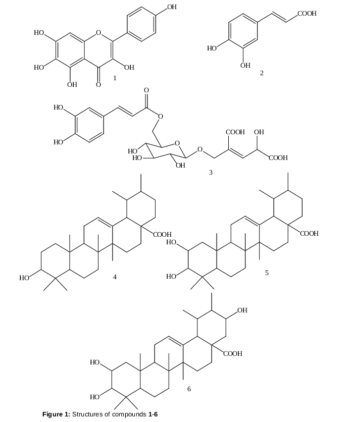
The chloroform soluble fraction of the methanol extract of the leaves of A. triflora was subjected to a series of column chromatographic separations. Three compounds, namely, ursolic acid (4), 2, 3-dihydroxy ursolic acid (5) and 2, 3, 21-trihydroxy ursolic acid (6) were obtained (). To the best of our knowledge, this is the first report of the isolation of ursolic acid based triterpenes from genus Abelia. The structures of the isolated compounds were established by UV, IR, MS and NMR spectroscopy. Three of our previously isolated compounds, 5,6,7,4-tetrahydroxy flavone 1, caffeic acid 2, abeliaside 3 [4], and the three triterpenes (4-6) that were isolated in this study, were all subjected to the biological investigation.
Anti-inflammatory and antioxidant activities
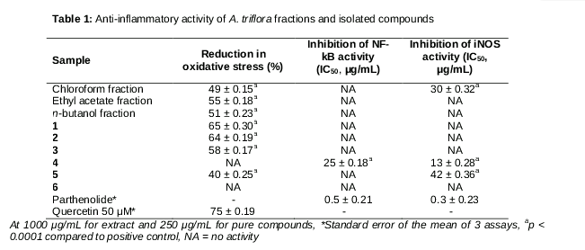
From the results seen in , the chloroform, ethyl acetate and n-butanol soluble fractions showed a decrease of 49, 55 and 51 % in the oxidative stress at 1000 µg/mL concentration in the cellular antioxidant assay using ABAP-induced HepG2 cells, respectively. Compounds 1-3 and 5 showed a decrease of 40 – 65 % in the oxidative stress at 250 µg/ml concentration, while compounds 4 and 6 were inactive.
The chloroform fraction and ursolic acid (4) inhibited iNOS activity with IC50 values of 30 and 13 µg/mL in lipopolysaccharide (LPS)-induced macrophages, while compounds1-3 were inactive and 5 was weakly active (IC50 42 µg/mL). Ursolic acid (4) was the only compound effective in inhibiting the NF-kB activity with an IC50 value of 25 µg/ml, compared to the positive standard Parthenolide. The NF-kB activity was lost in dihydroxy and trihydroxyursolic acids (5 and 6).
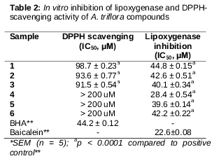
For the DPPH scavenging assay results shown in , compounds 1-3 exhibited moderate antioxidant activities compared with the positive control. As for the results of the lipoxygenase inhibitory effect seen in , compound 4 showed a potent inhibitory potential, while compounds 1-3 and 5-6 were moderate in activity.
Cytotoxic activity
Among the tested compounds only ursolic acid (4) exhibited cytotoxic activity against three human cancer cell lines including melanoma (SK-MEL), breast cancer (BT-549), and ovarian cancer (SK-OV-3) with IC50 values of 14.5, 16.0 and 12.5, respectively. The compound was also toxic to the two normal kidney cell lines, African Green Monkey kidney fibroblast (VERO) and pig kidney epithelial (LLC-PK1) cells with IC50 values 15.0 and 13.3, respectively. In contrast, 2,3-dihydroxy and 2,3,21-trihydroxyursolic acids were not cytotoxic to all tested cell lines.
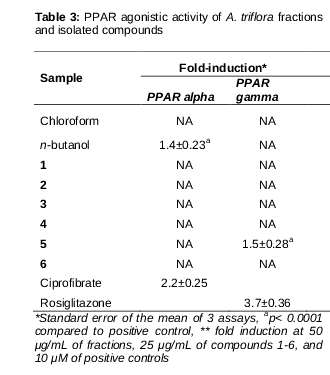
The peroxisome proliferator activated receptors (PPARα and PPARγ) are ligand dependent transcription factors that regulate the carbohydrate and lipid metabolism as well as inflammatory process. These are all considered significant targets related to metabolic disorder which is a combination of cardiovascular disease, diabetes, and inflammation [18]. Although ursolic acid (4) did not show any activation of PPARα or PPARγ, 2,3-dihydroxy ursolic acid (5) caused an increase of 1.5 fold in PPARγ activity at 25 µg/mL, compared to the positive standard, rosiglitazone ().
Discussion
The study resulted in the isolation of more bioactive metabolites (4-6) from A. triflora, which adds to the new reports on the plant. Results of the biological assays indicated that among the tested compounds, only ursolic acid (4) demonstrated anti-inflammatory activities in terms of multiple targets, contributing to the beneficial effects of this plant. Among the three tested triterpene acids (4-6), ursolic acid was found to be the most active as anti-inflammatory, which indicates that the hydroxylation at positions 2 and 21 seemed to weaken its activity. This is in agreement with previous literature which demonstrates the anti-inflammatory activity of ursolic acid [19,20]. Our results also proved that ursolic acid was the most active as cytotoxic constituent. This is in agreement with previous studies which confirmed the great potential of this triterpene acid as cytotoxic agent on different cell lines [21,22]. It also proves that the hydroxylation of ursolic acid at positions 2 and 21 may be responsible for lowering its anti-inflammatory and cytotoxic potential. This type of structure activity relationship is of utmost importance in natural product research. The plant could be also considered as a promising source of this valuable bioactive compound due to its high yield in the chloroform fraction. It is worth noting that ursolic acid did not show any activation of PPARα or PPARγ which are considered as targets for cardiovascular and diabetic therapy. However the cardiovascular effects of ursolic acids have been reported in literature [23]. Thus, our study proved that PPARα may not be involved in the cardiovascular action of ursolic acid. 2,3-dihydroxy ursolic acid (5) seemed to activate PPARγ without any effect on PPARα, which indicates its potential as antidiabetic drug. 2,3,21-trihydroxy ursolic acid (6) lost this PPAR agonistic activity, which proves the structure activity relationship between the number and position of hydroxyl groups on the triterpene skeleton and the PPAR agonistic activity. To the best of our knowledge, this is the first report on biological activities of these triterpene acids.
Conclusion
This study led to the isolation of more biologically active compounds from A. triflora than was previously done. Among the investigated compounds, ursolic acid was the most biologically active as an anti-inflammatory and cytotoxic drug. The 2,3-dihydroxyursolic acid shows promising anti-diabetic potential, which could open the way to the development of a new lead compound.
Declarations
Acknowledgement
References
Archives


News Updates